Bridge (dentistry)
| Bridge (dentistry) | |
|---|---|
| ICD-9-CM | 23.42-23.43 |
| MeSH | D003829 |
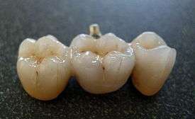

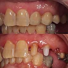
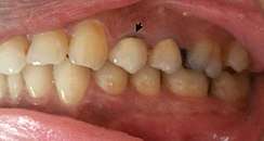
A bridge is a fixed dental restoration (a fixed dental prosthesis) used to replace one or more missing tooth by joining an artificial tooth definitively to adjacent teeth or dental implants.
A bridge will span the area where teeth are missing. They are attached to the natural teeth or implants that surround this space. The natural teeth or implants which support the bridge are called abutments. Depending on the type of bridge, natural abutment teeth may be reduced in size to accommodate the bridge to fit over them. An impression will be taken of the abutment tooth or implant and space to provide a mould to create the bridge. Using this, the bridge is then fabricated in a dental laboratory.
Definitions
Fixed bridge: A dental prosthesis that is definitively attached to natural teeth and replaces missing teeth.[1]
Abutment: The tooth that supports and retains a dental prosthesis.[2]
Pontic: The artificial tooth that replaces a missing natural tooth.[2]
Retainer: The component attached to the abutment for retention of the prosthesis.[2]
Unit: Pontics and abutment teeth are referred to as units. The total number of units in a bridge is equal to the number of pontics plus the number of abutment teeth.[1]
Case selection and treatment planning
Case selection
Appropriate case selection is important when considering the provision of fixed bridgework. Patient expectations should be discussed and a thorough patient history should be obtained. Replacement of missing teeth with fixed bridgework may not always be indicated and both patient factors alongside restorative factors should be considered before deciding if providing fixed bridgework is appropriate.[3] The survival rate of bridgework can be affected by the span of bridge needed, the proposed position of the bridge, and the size, shape, number and condition of planned abutment teeth.[4] Furthermore, any active disease including caries or periodontal disease should be treated and followed by a period of maintenance to ensure patient compliance in maintaining appropriate oral hygiene.[5][6]
Selection and evaluation of abutment teeth
Multiple factors influence the selection of appropriate abutment teeth. These include the size of potential abutment tooth, with larger teeth having an increased surface area preferable for retention, using teeth with a stable periodontal status, favourable tooth angulation, favourable tooth position, and an adequate crown-root ratio.[1][7]
Previously Ante's law, which states that the roots of abutment teeth must have a combined periodontal surface area in three dimensions that is more than that of the missing root structures of the teeth replaced with a bridge, was used in bridgework design. However, research has shown no evidence supporting Ante's law [8]
Indications for use
- Replacement of a single tooth or a small spanning space.
- Good oral health status and motivation of patient to maintain oral health.[9]
- Periodontal status of remaining dentition at a stable and satisfactory level.[9]
- Abutment teeth of good quality with minimal restorations and enough surface area and enamel present for adhesion.[7]
- Splinting of periodontally compromised teeth to improve occlusal stability, comfort and decrease mobility. (Periodontally compromised teeth is also a contraindication).[7]
- As a way of fixed retention after orthodontic treatment or extraction.[7]
- Patient unsuitable for implants. This may be due to inadequate bone levels, expense or patient not wanting to receive implants.[7]
Contraindications
- Size of saddle area too long.[10]
- Patients with parafunction e.g. bruxism.[10]
- Tooth mobility increases risk of de-bonding.[7]
- Malaligned teeth resulting in poor aesthetics and common path of insertion.[7]
- Abutment tooth quality inadequate for example may have a reduced surface area, reduced enamel or be heavily restored.[7]
- Increased risk of caries due to increased difficulty in maintaining oral hygiene around the bridgework.[7]
- Increased risk of loss of vitality.
- Allergy to base metal alloys e.g. nickel [7]
Types of bridge
Conventional bridge
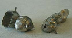
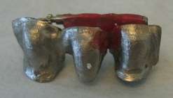
Conventional bridges are bridges that are supported by full coverage crowns, three-quarter crowns, post-retained crowns, onlays and inlays on the abutment teeth. In these types of bridges, the abutment teeth require preparation and reduction to support the prosthesis. Conventional bridges are named depending on the way the pontic (false teeth) is attached to the retainer.[11]
Fixed-fixed bridges
A fixed-fixed bridge refers to a pontic which is attached to a retainer at both sides of the space with only one path of insertion.
Cantilever
A cantilever is a bridge where a pontic is only attached to a retainer only at one side.
Spring cantilever
The pontic and retainer are remote from each other and connected by a metal bar. Usually, a missing anterior tooth is replaced and supported by a posterior tooth.
Fixed-movable
The pontic is firmly attached to a retainer at one end of the span (major retainer) and attached via a movable joint at the other end (minor retainer)
A major advantage of this type of bridge is that the movable joint can accommodate the angulation differences in the abutment teeth in long axis which enables the path of insertion to be irrespective of the alignment of the abutment tooth.[11]
Adhesive bridge
An alternative to the traditional bridge is the adhesive bridge (also called a Maryland bridge). An adhesive bridge utilizes "wings" on the sides of the pontic which attach it to the abutment teeth. Abutment teeth require minor or no preparation. They are most often used when the abutment teeth are whole and sound (i.e., no crowns or major fillings).
Types of artificial plastic teeth
Types of Artificial Plastic Teeth (known in the industry as Pontics)
- Wash-through Pontic[12]
- Dome Pontic (also known as bullet or torpedo shaped)[12]
- Ridge Lap Pontic (also known as Full Saddle Pontic)[12]
- Modified Ridge-Lap Pontic[12]
- Ovate Pontic[12] comes into contact with the underlying soft tissue and hides the defects of the edentulous ridge with applying light pressure.[13] It is commonly used in provisional bridges following extraction of teeth to improve the emergence profile and helps in shaping the gingiva around the future fixed prosthesis.[14]
Types of bridges according to durability
Bridges can either be provisional (temporary / interim) or permanent. The provisional bridge is a transitional restoration that protects the teeth that are weakened by the preparation, and stabilizes the dental tissues till the fabrication of the final restoration, moreover, it can pave the way to the esthetics of the future permanent restoration and its appearance, which can help the patient accept the final profile.[15] Provisional restorations are designed to be used for a few weeks to months, they can be fabricated directly (by chair side), or indirectly ( in the dental laboratory). It is usually tried in a few times to check if it fits properly and if its margins are well adapting on the teeth surface and gingiva, it may need relining or a few adjustments.[16] Provisional bridges can either be made of acrylic resins or metal. The resins are the most commonly used, they are either made of cellulose acetate, polycarbonate or poly-methyl methacrylate. Other chemically activated resins include poly-R methacrylates: these are methacrylates with ethyl or isobutyl substances added to increase the strength of material. Also, commonly used resins include the BisGMA based dimethacrylate, and the visible light urethane di-methylacrylate.[16][17] Dimethacrylate-based materials were found to be better than monomethacrylates for temporary restorations in terms of flexural strength and hardness.[18][19]
Types of bridges according to material
- Metal based, ex: noble based metals (Gold) , or base metal alloys, a.k.a. non-precious alloys (Nickle chromium)
- Non-metal based. They can be either resin veneered, fibre-reinforced composite, porcelain fused to metal, or ceramics which are either silica, alumina, or zirconia.[15]
Acrylic resin & Porcelain fused to metal (PFM)
Acrylic resin was the first veneering material used to help restore the aesthetics of crown and bridges, the aim was to maintain a similar colour to natural teeth by attaching it on the labial surface of metal crown / bridges, however, resin veneered dental prosthetics lacked stability and abrasion resistance.[15] Porcelain fused to metal (PFM) was then introduced; the porcelain is composed of two layers (one opaque to cover the metal substructure and another translucent to provide an enamel illusion). Still several researchers consider PFM the gold standard as it has been reported to have 95% success over a 10 year period, a reason why newer types of all-ceramic restorations are usually compared to PFM crowns / bridges to assess its success and durability.[20] However, PFM restorations may show a grey colour at the cervical margins of the tooth showing the metal substructure.
IPs Emax
IPs Emax ceramics offer high aesthetic properties, that's why its use has been increasingly popular, however, there's insufficient evidence to determine the longevity of Emax in bridges; some reports found fair short-term survival, but unfavorable medium-term survival.[21] Failures of restorations were most reported in the posterior teeth region. IPs Emax is available as press ingots or as IPs Emax CAD-CAM system.[22] Emax use in crowns or bridges is not recommended for patients who suffer from bruxism.[23]
Zirconia
Zirconia is used in anterior, and posterior fixed bridges, also on implants. Zirconia is fabricated using the dental CAD-CAM technology.[24] It has high mechanical strength and it can withstand high occlusal forces compared to all ceramic materials.[25] in addition it can resist crack propagation in the core material, however, cracks often occur in the veneering material leading to its fracture whether in the tooth supported or implant-supported bridges.[26][27] Reports found that the 3×3 mm designed connectors in zirconia bridges increased the strength to resist fracture by 20%.[28][29]
Although the use of ceramic based fixed prosthesis have been popular as it achieves a lifelike, highly esthetic appearance, a Cochrane review found insufficient evidence to support or refute the effectiveness of ceramic materials for fixed prosthodontic treatment over metal-ceramic.[30]
Clinical stages of bridgework
- Tooth preparation: This should be completed with reference to radiographs and study casts obtained during treatment planning. For conventional bridges, tooth preparation should aim to conserve tooth tissue, ensure a parallel path of insertion, achieve clearance in the occlusion and ensure well defined preparation margins.[9] The taper of each preparation on the abutment teeth must be the same. This is known as parallelism among the abutments and allows the bridge to fit onto the abutment teeth. Adhesive bridges require minimal preparation.
- Master impressions: An accurate impression should be made of the prepared teeth, along with an impression of the opposing arch. The master casts are used to provide accurate information about the occlusion to the laboratory and construct the prosthesis.[9]
- Occlusal registration: An occlusal registration is needed when providing extensive bridgework to allow the opposing casts to be related accurately. This may not be necessary if only a small number of teeth are to be restored.[9]
- Temporary restoration: Temporary restorations should be fabricated if possible to protect and maintain the prepared teeth until placement of the final restoration.[9]
- Try in: Confirm the clinical acceptability before cementing definitively. Assess the prosthesis on the master casts and identify the cause of any problems if present. A period of temporary cementation to assess clinical acceptability prior to definitive placement is sometimes used.[9]
- Final placement: Once satisfied the prosthesis is clinically acceptable, cement and bond the bridgework definitively.[9]
- Review: Assess the bridgework and manage any post-operative issues.[9]
Restoration fabrication
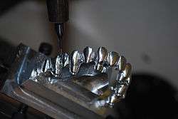
As with single-unit crowns, bridges may be fabricated using the lost-wax technique if the restoration is to be either a multiple-unit FGC or PFM. Another fabrication technique is to use CAD/CAM software to machine the bridge.[31] As mentioned above, there are special considerations when preparing for a multiple-unit restoration in that the relationship between the two or more abutments must be maintained in the restoration. That is, there must be proper parallelism for the bridge to seat properly on the margins.
Sometimes, the bridge does not seat, but the dentist is unsure whether it is because the spatial relationship between the abutments is incorrect, or whether the abutments do not actually fit the preparations. The only way to determine this is to section the bridge and try in each abutment by itself. If they each fit individually, the spatial relationship was incorrect, and the abutment that was sectioned from the pontic must now be reattached to the pontic according to the newly confirmed spatial relationship. This is accomplished with a solder index.
The proximal surfaces of the sectioned units (that is, the adjacent surfaces of the metal at the cut) are roughened and the relationship is preserved with a material that will hold on to both sides, such as GC pattern resin. With the two bridge abutments individually seated on their prepared abutment teeth, the resin is applied to the location of the sectioning to reestablish a proper spatial relationship between the two pieces. This can then be sent to the lab where the two pieces will be soldered and returned for another try-in or final cementation.
Advantages of bridges
Dental bridges offer several advantages.
They can usually be completed in only two dental appointments, restore the tooth back to full chewing function, require no periodic removal for cleaning, have a long life-expectancy and are aesthetically pleasing.[32]
Bridge failures
Common reasons for bridge failures
- Poor oral hygiene: As with any fixed prosthesis including bridges, maintaining good oral hygiene to prevent plaque formation around the bridge is key. This will ensure prolonged performance. A study examined the gingival health around the fixed bridges after a 14 day - 6 month post insertion found the surfaces were more plaque retentive, causing gingival inflammation regardless the material of fabrication of the bridge, unlike single crowns which didn't show the same effect.[33][34]
- Mechanical failures: These failures can occur due to loss of retention of the bridge due to improper cementation, construction or preparation.[35][36]
Fracture of the metal coating or pontic can also lead to mechanical failures. Fracture in connectors of bridges at the gingival side is a common finding in most all-ceram bridges.[37]
- Biological failures: These can occur due to caries in the tooth (one of the commonest causes of crown and bridge failures[6]) or due to pulpal injury. Problems with abutment teeth such as tooth fracture, secondary caries or periodontal disease can cause discomfort and put pressure on surrounding soft tissues to also cause a biological failure of the bridge.
- Aesthetic failures: These can occur at the time of cementation and include; colour mismatch, roughness of margins or improper tooth contour.
Aesthetics failures can also occur over a period of time including through wear of teeth, gingival recession or drifting of teeth.
- Problems with abutment teeth: Abutment teeth affected by secondary caries, vitality loss or periodontal disease can all lead to bridge failure.[35]
Oral manifestations of bridge failures
Bridge failures result in clinical complications and patients can present with:
- Pain in the oral cavity
- Sensitivity, bleeding and inflammation of the gums [35]
- Foul breath and taste disturbances
Bridge failure management
Management of bridge failures depend upon the extent and type of failure and these can be prevented through forming a thorough treatment plan with the patient as well regularly emphasising the importance of maintaining a very good level of oral hygiene after the bridge has been placed. The importance of cleaning underneath the pontic, through the use of interdental cleaning aids, should also be reinforced as plaque control around fixed restorations is more difficult.[6]
Management options include:
- Keeping the bridge under observation/review
- Repairing, replacing or removing the fault [6]
See also
References
- 1 2 3 Mitchell DA, Mitchell L, McCaul L (2014). Oxford Handbook of Clinical Dentistry (Sixth ed.). Oxford: Oxford University Press. p. 268.
- 1 2 3 "The Glossary of Prosthodontic Terms: Ninth Edition". The Journal of Prosthetic Dentistry. 117 (5S): e1–e105. May 2017. doi:10.1016/j.prosdent.2016.12.001. PMID 28418832.
- ↑ Hemmings K, Harrington Z (April 2004). "Replacement of missing teeth with fixed prostheses". Dental Update. 31 (3): 137–41. doi:10.12968/denu.2004.31.3.137. PMID 15116483.
- ↑ Bishop K, Addy L, Knox J (2007). "Modern restorative management of patients with congenitally missing teeth: 3. Conventional restorative options and considerations". Dental Update. 34 (1): 30–2, 34, 37–8. doi:10.12968/denu.2007.34.1.30. PMID 17348556.
- ↑ Maglad AS, Wassell RW, Barclay SC, Walls AW (August 2010). "Risk management in clinical practice. Part 3. Crowns and bridges". British Dental Journal. 209 (3): 115–22. doi:10.1038/sj.bdj.2010.675. PMID 20706245.
- 1 2 3 4 Briggs P, Ray-Chaudhuri A, Shah K (March 2012). "Avoiding and managing the failure of conventional crowns and bridges". Dental Update. 39 (2): 78–80, 82–4. doi:10.12968/denu.2012.39.2.78. PMID 22482265.
- 1 2 3 4 5 6 7 8 9 10 Gulati JS, Tabiat-Pour S, Watkins S, Banerjee A (2016). "Resin-Bonded Bridges – the Problem or the Solution? Part 1- Assessment and Design". Dental Update. 43 (6): 506–8, 510–2, 515–8, 521. doi:10.12968/denu.2016.43.6.506. PMID 29148644.
- ↑ Lulic M, Brägger U, Lang NP, Zwahlen M, Salvi GE (June 2007). "Ante's (1926) law revisited: a systematic review on survival rates and complications of fixed dental prostheses (FDPs) on severely reduced periodontal tissue support". Clinical Oral Implants Research. 18 Suppl 3: 63–72. doi:10.1111/j.1600-0501.2007.01438.x. PMID 17594371.
- 1 2 3 4 5 6 7 8 9 Ibbetson R, Hemmings K, Harris I (May 2017). "Guidelines for Crowns, Fixed Bridges and Implants". Dental Update. 44 (5): 374–6, 378–80, 382–4, 386. doi:10.12968/denu.2017.44.5.374. PMID 29188690.
- 1 2 Dayanik S (April 2016). "Resin-Bonded Bridges--Can We Cement Them 'High'?". Dental Update. 43 (3): 243–4, 247–50, 253. doi:10.12968/denu.2016.43.3.243. PMID 27439271.
- 1 2 Bartlett D, Ricketts D (2013). Advanced operative dentistry : a practical approach. Edinburgh: Elsevier Churchill Lvgst. ISBN 978-0-7020-5538-6.
- 1 2 3 4 5 Gopakumar A, Boyle EL (September 2013). "'A bridge too far'--the negative impact of a bridge prosthesis on gingival health and its conservative management". British Dental Journal. 215 (6): 273–6. doi:10.1038/sj.bdj.2013.877. PMID 24072295.
- ↑ Reddy K, Hegde V, Aparna IN, Dhanasekar B (2009). "Incorporating modified ovate pontic design for anterior tooth replacement: A report of two cases". The Journal of Indian Prosthodontic Society. 9 (2): 100–4. doi:10.4103/0972-4052.55254.
- ↑ network, MEFANET, Czech and Slovak medical faculties. "Division of pontics according to shape and relation to the mucosa. - WikiLectures". www.wikilectures.eu. Retrieved 2018-06-14.
- 1 2 3 Rosenstiel SF, Land MF, Junhei F (2006). Contemporary fixed prosthodontics (4th ed.). St. Louis: Mosby. ISBN 978-0-323-02874-5.
- 1 2 Smith BG (1998). Planning and Making Crowns and Bridges (3rd ed.). London: Taylor & Francis, A Martin Dunitz Book. ISBN 978-0-203-41955-7.
- ↑ Nallaswamy D (2003). Textbook of prosthodontics (1st ed.). New Delhi: Jaypee Brothers Medical Publishers. ISBN 9788180611995.
- ↑ Kim SH, Watts DC (January 2004). "Polymerization shrinkage-strain kinetics of temporary crown and bridge materials". Dental Materials. 20 (1): 88–95. doi:10.1016/S0109-5641(03)00101-5. PMID 14698778.
- ↑ Astudillo-Rubio D, Delgado-Gaete A, Bellot-Arcís C, Montiel-Company JM, Pascual-Moscardó A, Almerich-Silla JM (2018-02-28). "Mechanical properties of provisional dental materials: A systematic review and meta-analysis". PLOS One. 13 (2): e0193162. doi:10.1371/journal.pone.0193162. PMC 5830998. PMID 29489883.
- ↑ Ahmad I (2012). Prosthodontics at a glance. Chichester, West Sussex, UK: Wiley-Blackwell. ISBN 978-1-405-17691-0.
- ↑ Pieger S, Salman A, Bidra AS (July 2014). "Clinical outcomes of lithium disilicate single crowns and partial fixed dental prostheses: a systematic review". The Journal of Prosthetic Dentistry. 112 (1): 22–30. doi:10.1016/j.prosdent.2014.01.005. PMID 24674802.
- ↑ Willard A, Gabriel Chu TM (April 2018). "The science and application of IPS e.Max dental ceramic". The Kaohsiung Journal of Medical Sciences. 34 (4): 238–242. doi:10.1016/j.kjms.2018.01.012. PMID 29655413.
- ↑ Magne P, Perroud R, Hodges JS, Belser UC (October 2000). "Clinical performance of novel-design porcelain veneers for the recovery of coronal volume and length". The International Journal of Periodontics & Restorative Dentistry. 20 (5): 440–57. PMID 11203582.
- ↑ Manicone PF, Rossi Iommetti P, Raffaelli L (November 2007). "An overview of zirconia ceramics: basic properties and clinical applications". Journal of Dentistry. 35 (11): 819–26. doi:10.1016/j.jdent.2007.07.008. PMID 17825465.
- ↑ Daou EE (2014). "The zirconia ceramic: strengths and weaknesses". The Open Dentistry Journal. 8: 33–42. doi:10.2174/1874210601408010033. PMC 4026739. PMID 24851138.
- ↑ Le M, Papia E, Larsson C (June 2015). "The clinical success of tooth- and implant-supported zirconia-based fixed dental prostheses. A systematic review". Journal of Oral Rehabilitation. 42 (6): 467–80. doi:10.1111/joor.12272. PMID 25580846.
- ↑ Larsson C (2011). "Zirconium dioxide based dental restorations. Studies on clinical performance and fracture behaviour". Swedish Dental Journal. Supplement (213): 9–84. PMID 21919311.
- ↑ Bahat Z, Mahmood DJ, Vult von Steyern P (2009). "Fracture strength of three-unit fixed partial denture cores (Y-TZP) with different connector dimension and design". Swedish Dental Journal. 33 (3): 149–59. PMID 19994565.
- ↑ Vult von Steyern P (2005). "All-ceramic fixed partial dentures. Studies on aluminum oxide- and zirconium dioxide-based ceramic systems". Swedish Dental Journal. Supplement (173): 1–69. hdl:2043/1635. PMID 16001730.
- ↑ Poggio CE, Ercoli C, Rispoli L, Maiorana C, Esposito M (December 2017). "Metal-free materials for fixed prosthodontic restorations". The Cochrane Database of Systematic Reviews. 12: CD009606. doi:10.1002/14651858.CD009606.pub2. PMID 29261853.
- ↑ WorkNC Dental machining video, “Dental Bridge implant CNC Machining 5 axis”
- ↑ "Dental bridges. Advantages and disadvantages". infodentis.com.
- ↑ Kc Basnyat S, Sapkota B, Shrestha S (October 2015). "Oral Hygiene and Gingival Health in Patients with Fixed Prosthodontic Appliances - A Six Month Follow-up". Kathmandu University Medical Journal. 13 (52): 328–32. PMID 27423283.
- ↑ Ortolan SM, Viskić J, Stefancić S, Sitar KR, Vojvodić D, Mehulić K (March 2012). "Oral hygiene and gingival health in patients with fixed prosthodontic appliances--a 12-month follow-up". Collegium Antropologicum. 36 (1): 213–20. PMID 22816223.
- 1 2 3 Mitchell L, Mitchell DA, McCaul L (2014). Oxford Handbook Clinical Dentistry (Sixth ed.). Oxford: Oxford University Press. p. 276.
- ↑ Hargreaves K, Berman L (2010). Cohen's Pathways of the Pulp Expert Consult. ISBN 978-0-323-06489-7.
- ↑ Plengsombut K, Brewer JD, Monaco EA, Davis EL (March 2009). "Effect of two connector designs on the fracture resistance of all-ceramic core materials for fixed dental prostheses". The Journal of Prosthetic Dentistry. 101 (3): 166–73. doi:10.1016/S0022-3913(09)60022-6. PMID 19231568.