Bowen's disease
| Bowen's disease | |
|---|---|
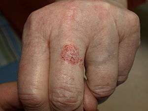 | |
| Bowen's disease as seen on a finger | |
| Specialty |
Oncology, Dermatology |
Bowen's disease, also known as squamous cell carcinoma in situ[1] is a neoplastic skin disease. It can be considered as an early stage or intraepidermal form of squamous cell carcinoma. It was named after John T. Bowen.
Erythroplasia of Queyrat is a particular type of Bowen's disease that can arise on the glans or prepuce in males, and, on the vulva in females, and may be induced by human papilloma virus.[2] It is reported to occur in the corneoscleral limbus.[3]
Signs and symptoms
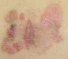
Bowen's disease typically presents as a gradually enlarging, well-demarcated red colored plaque with an irregular border and surface crusting or scaling. Bowen's disease may occur at any age in adults, but is rare before the age of 30 years; most patients are aged over 60. Any site may be affected, although involvement of palms or soles is uncommon. Bowen's disease occurs predominantly in women (70–85% of cases). About 60–85% of patients have lesions on the lower leg, usually in previously or presently sun-exposed areas of skin.
This is a persistent, progressive, unelevated, red, scaly or crusted plaque which is due to an intraepidermal carcinoma and is potentially malignant. The lesions may occur anywhere on the skin surface, including on mucosal surfaces. Freezing, cauterization, or diathermy coagulation is often effective treatment. Pathomorphologic study of tissue sampling revealed: polymorphism of spiny epithelial cells has progressed into atypism; increased mitosis; giant and multinucleate cells; acanthosis; hyperkeratosis and parakeratosis; basal membrane and basal layer are retained.
Causes
Causes of Bowen's disease include solar damage, arsenic, immunosuppression (including AIDS), viral infection (human papillomavirus or HPV), chronic skin injury, and other dermatoses.[4]
Histology
In Bowen's disease, atypical squamous cells proliferate through the whole thickness of the epidermis. The entire tumor is confined to the epidermis and does not invade into the dermis. The cells in Bowen's disease are often highly atypical under the microscope, and may in fact look more unusual than the cells of some invasive squamous cell carcinomas.
.jpg) Bowen's disease as seen under a microscope
Bowen's disease as seen under a microscope.jpg)
.jpg)
.jpg)
Treatment
Treatment options for Bowen's disease include photodynamic therapy with 5-aminolevulinic acid, cryotherapy, topical 5-fluorouracil or imiquimod, and excision. A meta-analysis showed evidence that PDL is more effective than cryotherapy and has better cosmetic outcomes. There is generally a lack of evidence comparing the effectiveness of all treatment options.[5]
References
- ↑ James, William; Berger, Timothy; Elston, Dirk (2005). Andrews' Diseases of the Skin: Clinical Dermatology. (10th ed.). Saunders. ISBN 0-7216-2921-0.
- ↑ J Invest Dermatol 2000 Sep;115(3):396–401
- ↑ Syed Imtiaz Ali Shah, Saeed Ahmed Sangi, Seraj Ahmed Abbasi: Bowen’s Disease: Pak J Ophthalmol 1998 Vol. 14, No. 1, pp. 37–38.
- ↑ Duncan KO; Geisse JK & DJ Leffell (2008). "Ch. 113: Epithelial precancerous lesions". In Wolff K; et al. Fitzpatrick's Dermatology in General Medicine (7th ed.). New York: McGraw-Hill. ISBN 978-0-07-146690-5.
- ↑ Bath-Hextall, Fiona J; Matin, Rubeta N; Wilkinson, David; Leonardi-Bee, Jo (2013-06-24). "Interventions for cutaneous Bowen's disease". Cochrane Database of Systematic Reviews. doi:10.1002/14651858.CD007281.pub2. ISSN 1465-1858.
External links
| Classification | |
|---|---|
| External resources |
| Wikimedia Commons has media related to Bowen's disease. |