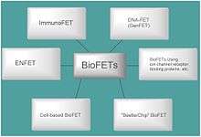
Bio-FET
Field-effect transistor-based biosensor (Bio-FET or BioFET) is a field-effect transistor that is gated by changes in the surface potential induced by the binding of molecules. When charged molecules, such as biomolecules, bind to the FET gate, which is usually a dielectric material, they can change the charge distribution of the underlying semiconductor material resulting in a change in conductance of the FET channel.[1][2][3] A Bio-FET consists of two main compartments: one is the biological recognition element and the other is the field-effect transistor.[4]

Mechanism of Operation
Bio-FETs couple a transistor device with a bio-sensitive layer that can specifically detect bio-molecules such as nucleic acids and proteins. A Bio-FET system consists of a semiconducting field-effect transistor that acts as a transducer separated by an insulator layer (e.g. SiO2) from the biological recognition element (e.g. receptors or probe molecules) which are selective to the target molecule called analyte.[6] Once the analyte binds to the recognition element, the charge distribution at the surface changes with a corresponding change in the electrostatic surface potential of the semiconductor. This change in the surface potential of the semiconductor acts like a gate voltage would in a traditional MOSFET, i.e. changing the amount of current that can flow between the source and drain electrodes.[7] This change in current (or conductance) can be measured, thus the binding of the analyte can be detected. The precise relationship between the current and analyte concentration depends upon the region of transistor operation.[8]
Fabrication of Bio-FET
The fabrication of Bio-FET system consists of several steps as follows:
- Finding a substrate suitable for serving as a FET site, and forming a FET on the substrate,
- Exposing an active site of the FET from the substrate,
- Providing a sensing film layer on active site of FET,
- Providing a receptor on the sensing film layer in order to be used for ion detection,
- Removing a semiconductor layer, and thinning a dielectric layer,
- Etching the remaining portion of the dielectric layer to expose an active site of the FET,
- Removing the photoresist, and depositing a sensing film layer followed by formation of a photoresist pattern on the sensing film,
- Etching the unprotected portion of the sensing film layer, and removing the photoresist[9]
Advantages
The principle of operation of Bio-FET devices based on detecting changes in electrostatic potential due to binding of analyte. This the same mechanism of operation as glass electrode sensors which also detect changes in surface potential but were developed as early as the 1920s. Due to the small magnitude of the changes in surface potential upon binding of biomolecules or changing pH, glass electrodes require a high impedance amplifier which increases the size and cost of the device. In contrast, the advantage of Bio-FET devices is that they operate as an intrinsic amplifier, converting small changes in surface potential to large changes in current (through the transistor component) without the need for additional circuitry. This means BioFETs have the capability to be much smaller and more affordable than glass electrode-based biosensors. If the transistor is operated in the subthreshold region, then an exponential increase in current is expected for a unit change in surface potential.
Bio-FETs can be used for detection in fields such as medical diagnostics,[10][9] biological research, environmental protection and food analysis. Conventional measurements like optical, spectrometric, electrochemical, and SPR measurements can also be used to analyze biological molecules. Nevertheless, these conventional methods are relatively time-consuming and expensive, involving multi-stage processes and also not compatible to real-time monitoring,[11] in contrast to Bio-FETs. Bio-FETs are low weight, low cost of mass production, small size and compatible with commercial planar processes for large-scale circuitry. They can be easily integrated into digital microfluidic devices for Lab-on-a-chip. For example, a microfluidic device in which controls sample droplet transport whilst enabling detection of bio-molecules, signal processing, and the data transmission, using an all-in-one chip.[12] Bio-FET also does not require any labeling step,[11] and simply utilise a specific molecular (e.g. antibody, ssDNA[13]) on the sensor surface to provide selectivity. Some Bio-FETs display fascinating electronic and optical properties. An example FET would is a glucose-sensitive based on the modification of the gate surface of ISFET with SiO2 nanoparticles and the enzyme glucose oxidase (GOD); this device showed obviously enhanced sensitivity and extended lifetime compared with that without SiO2 nanoparticles.[14]
Optimization
The choice of reference electrode (liquid gate) or back-gate voltage determines the carrier concentration within the field effect transistor, and therefore its region of operation, therefore the response of the device can be optimised by tuning the gate voltage. If the transistor is operated in the subthreshold region then an exponential increase in current is expected for a unit change in surface potential. The response is often reported as the change in current on analyte binding divided by the initial current ( ), and this value is always maximal in the subthreshold region of operation due to this exponential amplification.[8][15][16][17] For most devices, optimum signal-to-noise, defined as change in current divided by the baseline noise, ( ) is also obtained when operating in the subthreshold region,[8][18] however as the noise sources vary between devices, this is device dependent.[19]


One optimization of Bio-FET may be to put a hydrophobic passivation surface on the source and the drain to reduce non-specific biomolecular binding to regions which are not the sensing-surface.[20][21] Many other optimisation strategies have been reviewed in the literature.[8][22][23]
References
- ↑ Brand U, Brandes L, Koch V, Kullik T, Reinhardt B, Rüther F, Scheper T, Schügerl K, Wang S, Wu X: Monitoring and control of biotechnological production processes by Bio-FET-FIA-sensors, Appl Microbiol Biotechnol., 1991 Nov;36(2):167-72.
- ↑ Francesco Maddalena, Marjon J. Kuiper, Bert Poolman, Frank Brouwer, Jan C. Hummelen, Dago M. de Leeuw, Bert De Boer, and Paul W. M. Blom: Organic field-effect transistor-based biosensors functionalized with protein receptors, Journal of Applied Physics 108, 124501 (2010).
- ↑ Lin, M. C.; Chu, C. J.; Tsai, L. C.; Lin, H. Y.; Wu, C. S.; Wu, Y. P.; Wu, Y. N.; Shieh, D. B.; Su, Y. W. "Control and Detection of Organosilane Polarization on Nanowire Field-Effect Transistors". Nano Letters. 7 (12): 3656–3661. doi:10.1021/nl0719170.
- ↑ Joonhyung Lee, Piyush Dak, Yeonsung Lee, Heekyeong Park, Woong Choi, Muhammad A. Alam, Sunkook Kim: Two-dimensional Layered MoS2 Biosensors Enable Highly Sensitive Detection of Biomolecules, Sci. Rep., 2014; 4: 7352.
- 1 2 Schöning, Michael J.; Poghossian, Arshak (2002). "Recent advances in biologically sensitive field-effect transistors (BioFETs)". The Analyst. 127 (9): 1137–1151. doi:10.1039/B204444G. ISSN 0003-2654.
- ↑ Alena Bulyha, Clemens Heitzinger and Norbert J Mauser: Bio-Sensors: Modelling and Simulation of Biologically Sensitive Field-Effect-Transistors, ERCIM News, 04,2011.
- ↑ Matsumoto, A; Miyahara, Y (21 November 2013). "Current and emerging challenges of field effect transistor based bio-sensing". Nanoscale. 5 (22): 10702–10718. doi:10.1039/c3nr02703a.
- 1 2 3 4 Lowe, Benjamin M.; Sun, Kai; Zeimpekis, Ioannis; Skylaris, Chris-Kriton; Green, Nicolas G. (2017). "Field-effect sensors – from pH sensing to biosensing: sensitivity enhancement using streptavidin–biotin as a model system". The Analyst. Royal Society of Chemistry (RSC). 142 (22): 4173–4200. doi:10.1039/c7an00455a. ISSN 0003-2654.
- 1 2 Yuji Miyahara, Toshiya Sakata, Akira Matsumoto: Microbio genetic analysis based on Field Effect Transistors, Principles of Bacterial Detection: Biosensors, Recognition Receptors and Microsystems.
- ↑ Poghossian, A.; Cherstvy, A.; Ingebrandt, S.; Offenhäusser, A.; Schöning, M.J. (2005). "Possibilities and limitations of label-free detection of DNA hybridization with field-effect-based devices". Sensors and Actuators B: Chemical. Elsevier BV. 111-112: 470–480. doi:10.1016/j.snb.2005.03.083. ISSN 0925-4005.
- 1 2 K.Y.Park, M.S.Kim, K.M.Park, and S.Y.Choi: Fabrication of BioFET sensor for simultaneous detection of protein and DNA, Electrochem.org.
- ↑ Choi K, Kim JY, Ahn JH, Choi JM, Im M, Choi YK: Integration of field-effect transistor-based biosensors with a digital microfluidic device for a lab-on-a-chip application, Lab Chip., 2012 Apr
- ↑ Chu, Chia-Jung; Yeh, Chia-Sen; Liao, Chun-Kai; Tsai, Li-Chu; Huang, Chun-Ming; Lin, Hung-Yi; Shyue, Jing-Jong; Chen, Yit-Tsong; Chen, Chii-Dong. "Improving Nanowire Sensing Capability by Electrical Field Alignment of Surface Probing Molecules". Nano Letters. 13 (6): 2564–2569. doi:10.1021/nl400645j.
- ↑ Jing-Juan Xu, Xi-Liang Luo and Hong-Yuan Chen: ANALYTICAL ASPECTS OF FET-BASED BIOSENSORS, Frontiers in Bioscience, 10, 420--430, January 1, 2005
- ↑ Sarkar, Deblina; Liu, Wei; Xie, Xuejun; Anselmo, Aaron C.; Mitragotri, Samir; Banerjee, Kaustav (2014). "MoS2Field-Effect Transistor for Next-Generation Label-Free Biosensors". ACS Nano. 8 (4): 3992–4003. doi:10.1021/nn5009148. ISSN 1936-0851.
- ↑ Wen, Xuejin; Gupta, Samit; Nicholson, Theodore R.; Lee, Stephen C.; Lu, Wu (2011). "AlGaN/GaN HFET biosensors working at subthreshold regime for sensitivity enhancement". Physica Status Solidi C. 8 (7–8): 2489–2491. doi:10.1002/pssc.201001174. ISSN 1862-6351.
- ↑ Sun, K; Zeimpekis, I; Hu, C; Ditshego, N M J; Thomas, O; de Planque, M R R; Chong, H M H; Morgan, H; Ashburn, P (2016). "Effect of subthreshold slope on the sensitivity of nanoribbon sensors". Nanotechnology. 27 (28): 285501. doi:10.1088/0957-4484/27/28/285501. ISSN 0957-4484.
- ↑ Gao, Xuan P. A.; Zheng, Gengfeng; Lieber, Charles M. (2010). "Subthreshold Regime has the Optimal Sensitivity for Nanowire FET Biosensors". Nano Letters. 10 (2): 547–552. doi:10.1021/nl9034219. ISSN 1530-6984. PMC 2820132.
- ↑ Rajan, Nitin K.; Routenberg, David A.; Reed, Mark A. (2011). "Optimal signal-to-noise ratio for silicon nanowire biochemical sensors". Applied Physics Letters. 98 (26): 264107. doi:10.1063/1.3608155. ISSN 0003-6951. PMC 3144966.
- ↑ Kim JY, Choi K, Moon DI, Ahn JH, Park TJ, Lee SY, Choi YK: Surface engineering for enhancement of sensitivity in an underlap-FET biosensor by control of wettability, Biosens Bioelectron., 2013
- ↑ A. Finn, J.Alderman, J. Schweizer : TOWARDS AN OPTIMIZATION OF FET-BASED BIO-SENSORS, European Cells and Materials, Vol. 4. Suppl. 2, 2002 (pages 21-23)
- ↑ Schöning, Michael J.; Poghossian, Arshak (2002). "Recent advances in biologically sensitive field-effect transistors (BioFETs)". The Analyst. Royal Society of Chemistry (RSC). 127 (9): 1137–1151. doi:10.1039/b204444g. ISSN 0003-2654.
- ↑ Schöning, Michael J.; Poghossian, Arshak (2006). "Bio FEDs (Field-Effect Devices): State-of-the-Art and New Directions". Electroanalysis. Wiley-Blackwell. 18 (19–20): 1893–1900. doi:10.1002/elan.200603609. ISSN 1040-0397.