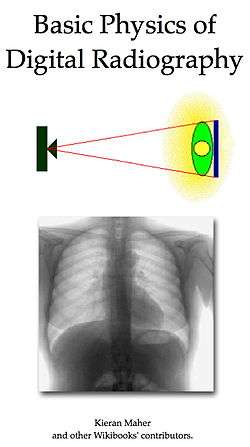
Digital Radiography heralds a new era for medical X-ray imaging. Radiographs can be recorded using digital image receptors and enhanced using computer processing. They can also be transferred to databases for archival and transmission throughout hospitals and clinics. The change from traditional film-based image receptors is similar in many respects to those occurring in digital photography and digital television. Greater precision can now be applied to each stage of the imaging process so that the balance between image quality and radiation dose can at last be controlled accurately. Knowledge and understanding of the basic physics which underlies practice is critical for its successful application in the clinical environment. This wikibook addresses this requirement.
The intent is a text which succinctly explains the physical basis of X-Rays and their modern application in Diagnostic Radiography. The wikibook is primarily for students with foundations in anatomy and physiology and could also be of interest to physics and engineering students requiring a topic overview. Math is kept to a minimum and only a basic knowledge of algebra is required of students. The use of calculus and other math techniques is largely avoided by using conceptual descriptions of the more complex processes. Keeping radiation dose as low as reasonably achievable while understanding its effects on image quality is the primary emphasis.
The focus extends from general radiography through fluoroscopy to 3D angiography as applied in general hospitals and clinics. The subject treatment is different from that used in conventional textbooks in that radiation biology is not treated as a separate topic but is embedded within the consideration of image formation. Likewise, the topic of image quality is integrated into the treatment of image display. The hope is that a more integrated treatment of the subject is developed for those studying the field for the first time.
The wikibook contains the following chapters:
- Atomic Structure || Electron Binding Energy || Electromagnetic Radiation || Diagnostic X-Rays || X-Ray Detection || Attenuation of X-Rays || Radiography Systems || The Fourier Transform
- X-Ray Tube || X-Ray Energy Spectrum || HV Generator || Beam Filtration || Light Beam Diaphragm || Automatic Exposure Control
- Interaction Processes || Radiation Biology || Tissue Attenuation || X-Ray Shadows || Subject Contrast || Radiation Dose || Radiation Protection
- Computed Radiography || Digital Radiography || XII-Video Systems || Radiation Dose Management
- Binary Representation || General-Purpose Computer || Digital Image Processor || Digital Images || Digital Image Processing || Image Storage & Distribution
- Image Display || Image Contrast || Spatial Resolution || Image Noise || Detective Quantum Efficiency || Temporal Resolution || Image Quality Assessment
- General Radiography || Digital Subtraction Angiography || Cone-Beam Computed Tomography || Dual-Energy Radiography || Image Superimposition
- X-ray Production & Interactions with Matter || Radiation Biology & Dosimetry || Technology of Radiographic Imaging
Further details are provided on The Contents page. An Index is also available. Note that the particular requirements of mammography and of paediatric, dental, chiropractic and veterinary radiography are not covered at this time.
A companion WikiBook on the Basic Physics of Nuclear Medicine is also available.
Principal Authors
As of February 2011, the principal author of this wikibook is Kieran Maher. You can send an e-mail message if you'd like to provide feedback, criticism, correction, additions/improvement suggestions etc. regarding this wikibook..... or even just to say hello!
Bibliography
- Bushberg JT, Seibert JA, Leidholdt & Boone JM, 2011. The Essential Physics of Medical Imaging, 3rd Ed..
- Gonzalez RC & Woods RE, 2008. Digital Image Processing, 3rd Ed..
- Heggie JCP, Liddell NA & Maher KP, 2001. Applied Imaging Technology, 4th Ed.
- Oppelt A, Ed., 2005. Imaging Systems for Medical Diagnostics.
External Links
Radiopaedia.org is a free educational radiology resource with one of the web's largest collections of radiology cases and reference articles.